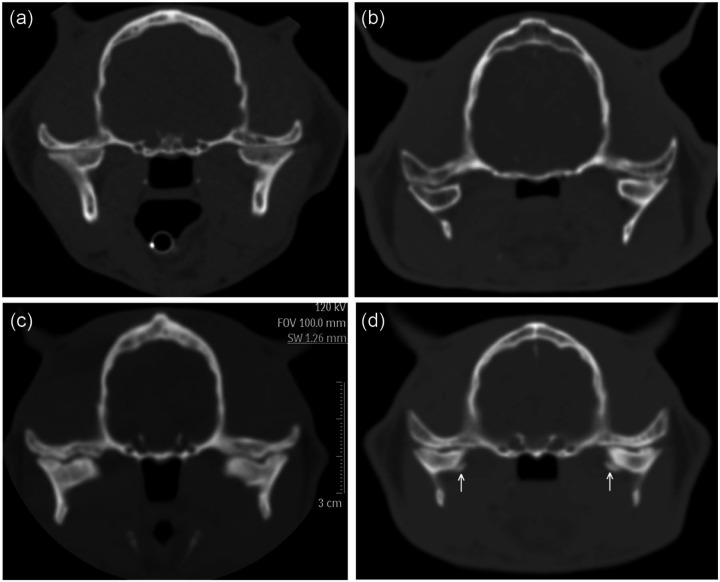
Figure 5.
Examples of temporomandibular joint (TMJ) malformation in cats with hypersomatotropism. (a) Transverse computed tomography (CT) image of a control cat with hypersomatotropism showing smooth straight surface of the mandibular condyles and narrow TMJ spaces. (b) Transverse CT image of a cat with hypersomatotropism showing concave surface of the mandibular condyles. The nasopharynx is markedly narrowed in this cat. (c) Transverse CT image of a cat with hypersomatotropism with symmetrical focal indentations in the mandibular condyles. The TMJ space is uneven in width. The parietal bone is thickened. (d) Transverse CT image of another cat with hypersomatotropism showing abnormal curvature of the mandibular condyles, widened TMJ space and bilateral angular osteophytes (arrows) affecting the medial aspects of the mandibular condyles

