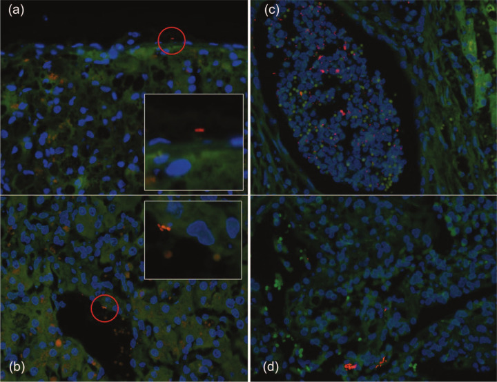
Figure 1.
Regional distribution of bacteria in hepatic biopsies. (a) A bacterium (EUB338-Cy3, red) is visualized on the capsule of the liver (magnified in insert). (b) Bacteria (Escherichia coli-Cy3,red) within a hepatic sinusoid (magnified in insert). (c) Multiple bacteria (EUB338-Cy3, red) within a hepatic micro-abscess. (d) Multiple bacteria (EUB338-Cy3, red) within the hepatic parenchyma

