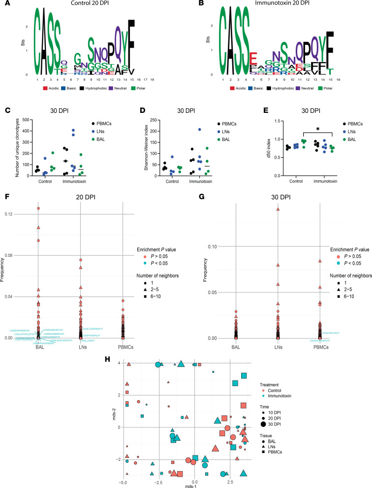
Figure 3. Immunotoxin administration does not modulate the clonotypic repertoire of CM9-specific CD8+ T cells during acute infection with SIV.
Experimental details as in Figure 2. (A) Logo plots and chemical classification of amino acids spanning the CDR3β loops of the top 10 pooled clonotypes from control macaques on day 20. (B) Logo plots and chemical classification of amino acids spanning the CDR3β loops of the top 10 pooled clonotypes from immunotoxin-treated macaques on day 20. (C) Repertoire diversity measured using the number of unique clonotypes for CM9-specific CD8+ T cell populations from control and immunotoxin-treated macaques on day 30. (D) Repertoire diversity measured using the Shannon-Weiner index for CM9-specific CD8+ T cell populations from control and immunotoxin-treated macaques on day 30. (E) Repertoire diversity measured using the d50 index for CM9-specific CD8+ T cell populations from control and immunotoxin-treated macaques on day 30. (F) TCRNET analysis of CM9-specific CD8+ T cell repertoires from control and immunotoxin-treated macaques on day 20. (G) TCRNET analysis of CM9-specific CD8+ T cell repertoires from control and immunotoxin-treated macaques on day 30. (H) Multidimensional scaling (MDS) analysis of CM9-specific CD8+ T cell repertoires from control and immunotoxin-treated macaques on days 10, 20, and 30. Clonotypes that were enriched in CM9-specific CD8+ T cell populations from immunotoxin-treated macaques are shown in blue. Each symbol represents 1 macaque (C–E and H). Horizontal bars indicate median values (C–E). Significance was determined using a 2-way ANOVA with Šídák correction (C–E). *P < 0.05. DPI, days postinfection.

