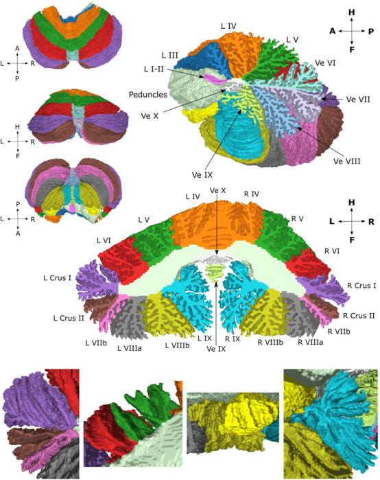
Figure 2.

Surface atlas from different views along with a sagittal and coronal cross-section that have been filled with the volumetric atlas. The bottom row shows enlarged images of the surface atlas from the anterior view of left crus I, II and lobule VIIb (left image), lobules V and VI (center-left image), lobules X and VIIIb (center-right image), and lobule IX (right image).
