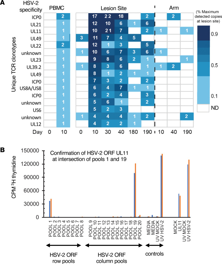
Figure 5. Representative data from fine specificity determination of blood CD4+ T cell clones overlapping with TCRβ CDR3 sequences detected in HSV lesion site biopsies.
(A) Clonotype tracking with assigned specificity of 13 of the 16 clones queried in P4 by abundance and fold change in skin over dose 1 (day 0–10). (B) Example of fine-specificity mapping of a single clone confirmed to be specific to UL11. Both specimens were obtained from day 10 after HSV529 vaccination. At left is T cell proliferation in response to matrix pools of HSV-2 antigens with positive and negative controls. Pools containing US11 (pool 1 and pool 19) are positive. At right is confirmatory assay with recombinant UL11 and controls. Blue and orange bars represent 2 replicate assays.

