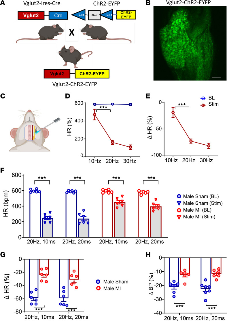
Figure 3. In vivo optogenetic stimulation of vagal sensory neurons in healthy control, sham, and infarcted male mice.
(A and B) Vglut2-ires-Cre mice were crossed with ChR2-EYFP mice to obtain Vglut2-ChR2-EYFP offspring, which was confirmed via genotype testing. In addition, presence of EYFP in the nodose ganglia of Vglut2-ChR2-EYFP mice was also confirmed via confocal microscopy. Scale bar: 50 µm. (C–E) In vivo responses to left vagal optogenetic stimulation in Vglut2-ChR2-EYFP mice are shown (n = 3). (F) Two to 3 weeks after MI, in vivo optogenetic stimulation of the left vagus nerve was performed, and hemodynamic responses were quantified at baseline and during stimulation. Both sham (n = 6) and infarcted male mice (n = 6) showed a decrease in HR in response to optogenetic stimulation (P < 0.0001). (G) However, changes in HR were greater in sham versus MI animals (20 Hz, 10 ms: male sham versus male MI, P < 0.0001; 10 Hz, 20 ms: male sham versus male MI, P < 0.0001). (H) Similarly, decreases in blood pressure to stimulation were greater in male sham versus MI animals (20 Hz, 10 ms: male sham (n = 7) versus male MI (n = 6), P < 0.0001; 20 Hz, 20 ms: male sham versus male MI, P < 0.0001). Data are shown as mean ± SEM. ***P < 0.001. BL, baseline prior to stimulation; Stim, during optical stimulation. Unpaired Student’s t test used for comparisons of MI versus sham.

