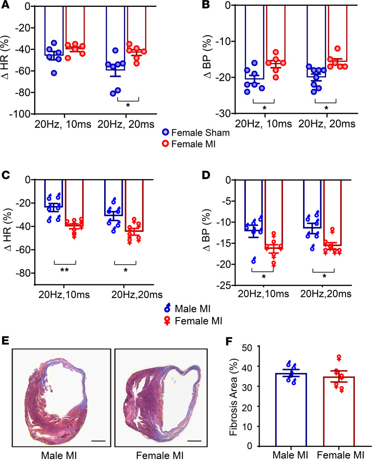
Figure 4. Sex differences in in vivo optogenetic responses.
(A and B) In response to in vivo optogenetic vagal stimulation, female MI animals demonstrated a blunted HR and BP response compared with female sham animals (n = 6 per group). (C) However, changes in HR were significantly different in male MI versus female MI animals (n = 6 per group) at both stimulation parameters (P < 0.01), with male animals demonstrating more diminished responses. (D) Similar to HR, blood pressure responses to optogenetic stimulation were also reduced in infarcted males versus infarcted females (male MI versus female MI change in blood pressure at 20 Hz, 10 ms: P < 0.05; change in blood pressure at 20 Hz, 20 ms: P < 0.05, n = 6 per group). (E and F) Myocardial fibrosis was quantified using Masson’s trichrome staining. Scale bar: 500 μm. No difference in the degree of fibrosis between male MI and female MI mice was observed (n = 5 per group). Data are shown as mean ± SEM. *P < 0.05, **P < 0.01. BL, baseline (prestimulation); Stim, during optical stimulation. Unpaired Student’s t test used for comparisons of males versus females.

