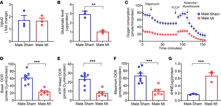
Figure 5. Vglut2 and glutamate levels, mitochondrial function, and oxidative stress in the vagal ganglia of male sham and infarcted mice.
(A and B) Vagal ganglia Vglut2 (n = 6 animals/sample, 3 separate samples per group) and glutamate levels (n = 9 animals/sample, 3 separate samples per group) were measured. While there was no change in Vglut2 levels, significantly lower glutamate levels (P < 0.01) were found in infarcted compared with sham males. (C) Mitochondrial oxygen consumption rates (OCRs) were measured in the vagal ganglia of sham (n = 8 ganglia) and infarcted (n = 6 ganglia) males. (D) Basal OCR was lower in infarcted versus sham males (P < 0.001). (E) ATP-linked OCR was also lower in infarcted versus sham males (P < 0.001). (F) Maximal OCR, deduced from treatment with FCCP (uncoupler), was found to be lower in infarcted versus sham males (P < 0.001). (G) 4-HNE levels were significantly lower in infarcted versus sham males (n = 6 animals/sample, 3 independent samples per group, P < 0.001). Data are shown as mean ± SEM. **P < 0.01, ***P < 0.001. Unpaired Student’s t test was used for intergroup comparisons.

