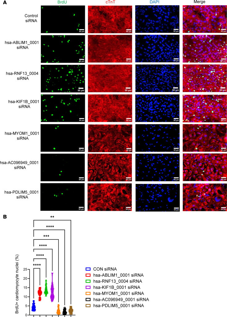
Figure 6. Evaluation of cardiomyocyte cell cycle via BrdU incorporation assay.
hiPSC-CMs at day 28 after initiation of cardiac differentiation were used. Cell cycle was assessed by immunostaining using antibodies against BrdU (to label cells in S phase). Cardiomyocytes were labeled using anti-human cardiac troponin T (cTnT) immunostaining; all nuclei were counterstained with DAPI. The prevalence of BrdU positively stained nuclei of hiPSC-CMs was counted and normalized to the total number of cardiomyocyte nuclei. Data were presented as percentage. (A) Representative images of anti-BrdU immunostaining in hiPSC-CMs. (B) Quantification of the prevalence of BrdU positively stained nuclei of hiPSC-CMs after siRNA-based knockdown. All data were presented as mean ± SEM. Statistical analysis was performed via 1-way ANOVA with Tukey’s honestly significant difference test. n = 30 technical replicates in each group. **P < 0.01, ***P < 0.001, and ****P < 0.0001.

