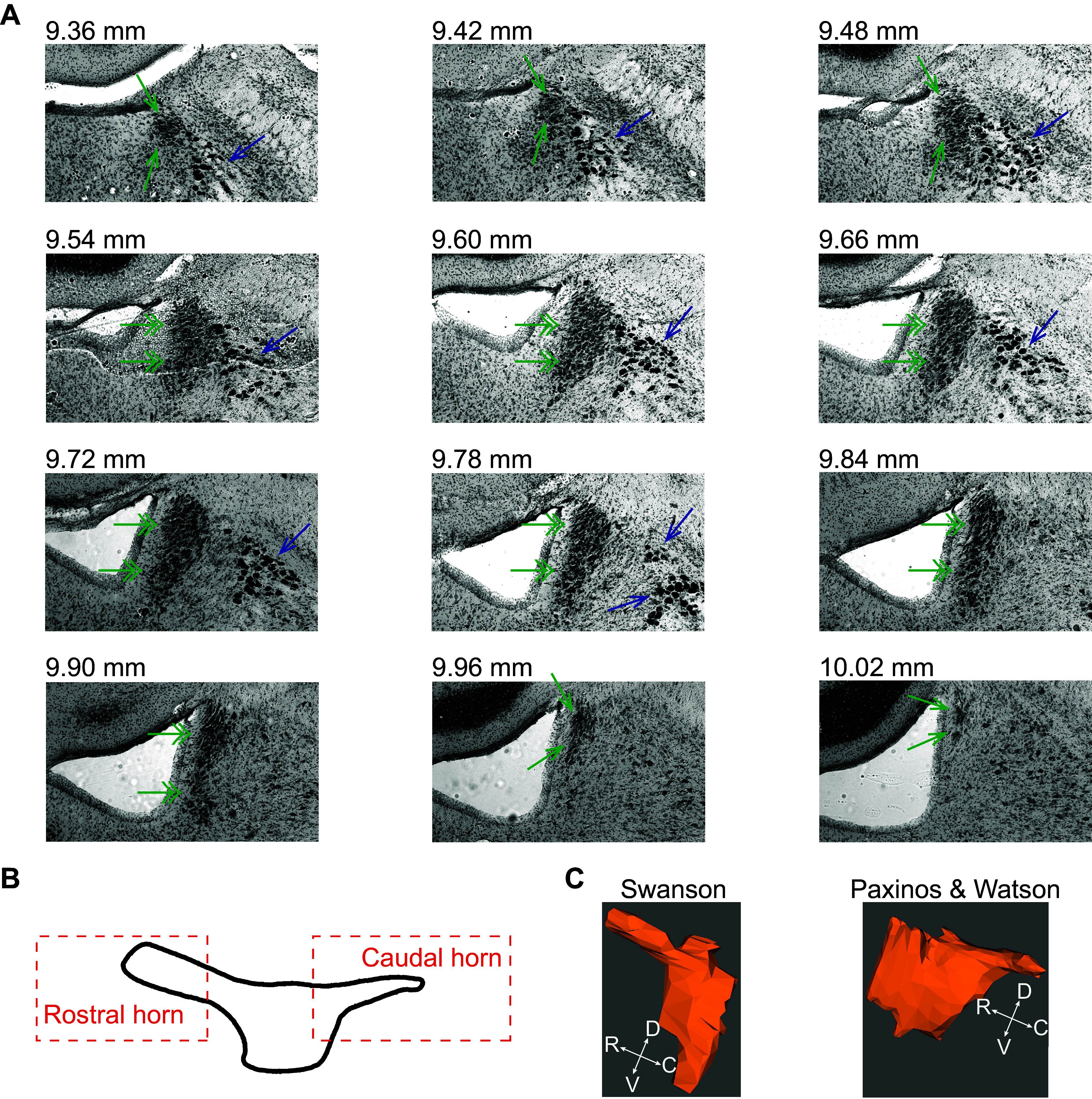
Figure 3.

The borders of the locus coeruleus (LC) central core and the rostral and caudal horns are cytoarchitectonically defined using a Nissl stain. A: the dense packing of LC neurons is shown from most rostral (top left) to most caudal (bottom right) in Nissl-stained frontal sections from the rat brain. The image is shown in grayscale. The numbers indicate rostrocaudal distance from Bregma. Single arrows in green indicate the LC horns, double arrows in green indicate the LC central core, and blue arrows indicate mesencephalic trigeminal nucleus (MeV). B: a reproduction of Loughlin’s depiction of the LC from a lateral view showing the central core and horns (67). C: two atlases of the rat brain differ in their depiction of the rostral and caudal aspects of the LC. Paxinos and Watson’s atlas shows a caudal horn but no rostral horn, whereas Swanson’s atlas shows a rostral horn but no caudal horn. The three-dimensional (3-D) renderings were constructed from coronal sections of each atlas for comparison using the neuroVIISAS platform (68, 69).
