Figure 4.
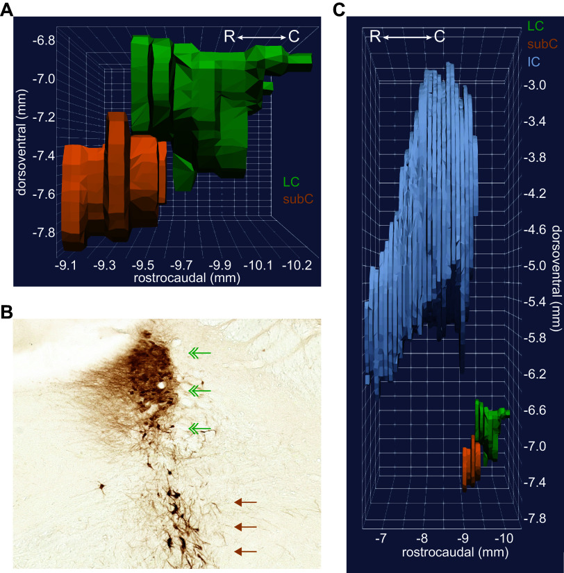
The subcoeruleus (SubC) is ventral to the rostral aspect of the locus coeruleus (LC) core and extends far rostral into coronal sections containing the inferior colliculus (IC). A: the three-dimensional (3-D) rendering shows the SubC region (orange) ventral to the LC core (green). The rendering was constructed from coronal sections in Paxinos and Watson’s rat brain atlas (70), with permission from Elsevier. B: a coronal section from the rat brain at the level of the rostral horn. Norepinephrine (NE) neurons appear as brown using a diaminobenzidine (DAB) and horse radish peroxidase reaction to visualize a tyrosine hydroxylase antibody. The rostral horn of the LC core is indicated by green double arrowheads. The SubC is indicated by brown single arrowheads. Note that SubC neurons are larger and arranged more diffusely compared with the LC core. This coronal section is from approximately −9.5 mm to −9.4 mm in the rostrocaudal plane, as shown in A. Note that the 3-D rendering in A does not appear to be a “horn” because that aspect of the LC is not contained within Paxinos and Watson’s rat brain atlas. C: the 3-D rendering shows the SubC extending rostrally into sections also containing the caudal aspects of the IC (gray), whereas LC neurons are not present in coronal sections containing the IC. Thus, the IC can be used in sagittal and coronal sections to determine whether NE neurons ventral to the 4th ventricle are LC core or SubC.

