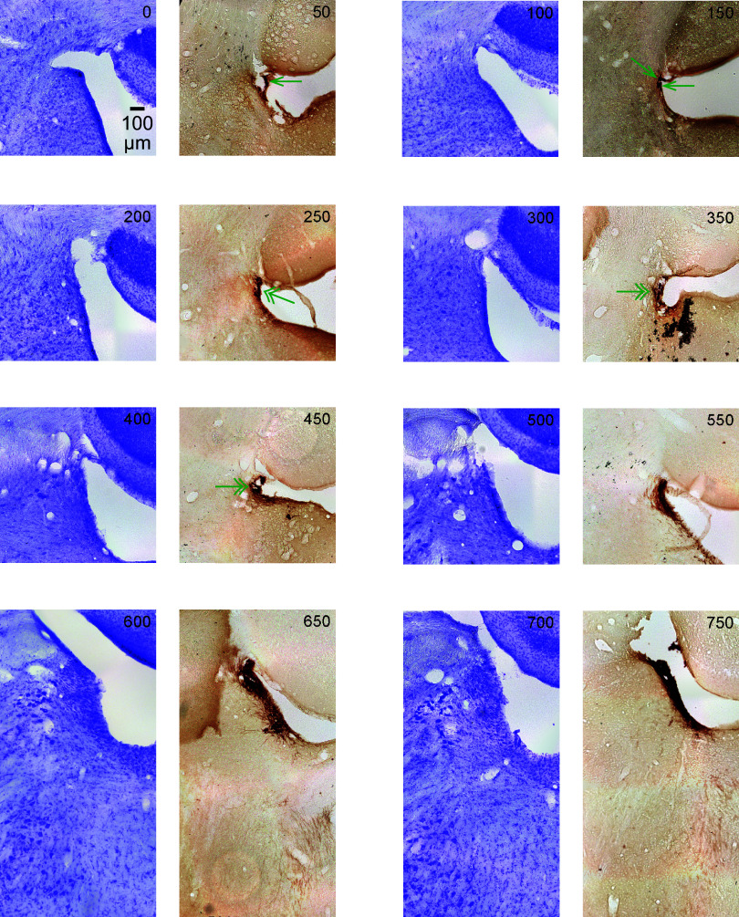
Figure 6.
The figure shows serial coronal sections of the rat (male) brainstem in 50-μm thickness. All sections are the same scale (see 100 μm scale bar). Upward is dorsal and leftward is lateral. The sections alternate Nissl (violet) and tyrosine hydroxylase (TH) immunostain (dark brown). This figure contains the most caudal sections, relative to Figs. 7, 8, and 9. The top left section is the most caudal section and is labeled 0. Subsequent serial sections are labeled in 50-μm increments from the first section and continue rostral (through Figs. 7, 8, and 9). In the first few caudal sections, where locus coeruleus (LC)-norepinephrine (NE) neurons are sparse, a single green arrowhead marks TH-positive neurons. In the next few sections, areas of increased density of TH-positive neurons are shown with double arrowheads. In subsequent sections, the TH-positive neurons are densely stained and are therefore left unmarked.

