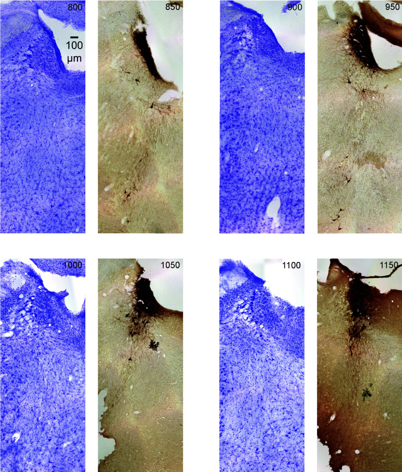
Figure 7.
The figure shows serial coronal sections of the rat (male) brainstem in 50-μm thickness continuing from Fig. 6. All sections are the same scale (see 100 μm scale bar). Upward is dorsal and leftward is lateral. The sections alternate Nissl (violet) and tyrosine hydroxylase (TH) immunostain (dark brown). The top left section is labeled in μm from the most caudal section in the top left corner of Fig. 6. Subsequent serial sections are labeled in 50-μm increments and continue rostral (through Figs. 8 and 9).

