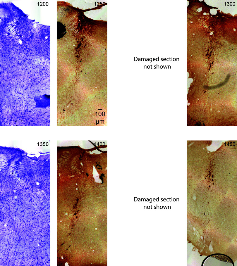
Figure 9.
The figure shows sections that are rostral to the locus coeruleus (LC) rostral horn (see Fig. 8. section marked 1250). Therefore, these sections run 250 μm to 600 μm rostral to the most rostral boundary of the LC. However, these sections still contain tyrosine hydroxylase (TH)-positive neurons, such as subcoeruleus (SubC)-norepinephrine (NE) neurons. Note that the section at 1650 contains an artifact, which is cerebellum tissue laying over the brainstem. The immunostained (dark brown) neurons are in the dorsal brainstem (SubC). The images are serial coronal sections of the rat (male) brainstem in 50-μm thickness continuing from Figs. 6, 7, and 8. All sections are the same scale (see 100 μm scale bar). Upward is dorsal and leftward is lateral. The sections alternate Nissl (violet) and TH immunostain (dark brown). The top left section is labeled in μm from the most caudal section in the upper left corner of Fig. 6. Subsequent serial sections are labeled in 50-μm increments and continue rostral (through to the bottom right of Fig. 9).

