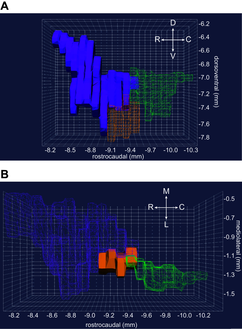
Figure 10.
The intermingling of the rostral locus coeruleus (LC) core, subcoeruleus (SubC), and laterodorsal tegmental nucleus (LDTg) presents potential for misclassifying GABAergic, glutamatergic, and cholinergic neurons as LC core neurons. A: the three-dimensional (3-D) rendering shows a lateral view of the LDTg (purple), SubC (orange), and LC (green). The SubC and LC are shown as wireframe renderings to highlight the overlap of the LTDg with norepinephrine (NE) neurons of the SubC and LC. In a sagittal section, the GABA, glutamate, and acetylcholine producing neurons of the LDTg can appear intermixed with NE neurons of the LC core. B: the 3-D rendering shows a dorsal view, looking ventrally into the pons. This view illustrates the close proximity of rostral LC core (green) and LDTg (purple). The LDTg and LC core are presented as wireframe renderings to emphasize that inserting a multi-electrode array near the rostral LC core with a slight angle can easily penetrate the LDTg, LC core, and SubC (orange) in a single-recording tract, or simultaneously record from LDTg on dorsal aspects of the array and SubC on ventral aspects of the array while missing the LC core altogether. This complication can be avoided when recording tracts are confined to the more caudal, central portion of the LC core (e.g., −9.7 to −10.1 mm on the rostrocaudal axis). In both panels, the 3-D renderings were constructed using coronal sections from Paxinos & Watson’s rat brain atlas [Paxinos and Watson (70), with permission from Elsevier].

