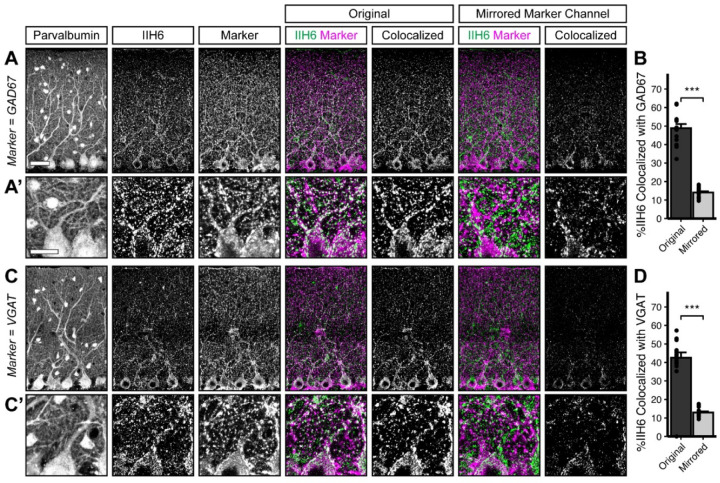
Figure 1. Dystroglycan co-localizes with markers of inhibitory synapses.
Cerebellar cortex of lobules V-VI were immunostained with Parvalbumin to show Purkinje cell and MLI morphology and counterstained with IIH6 (glycosylated Dystroglycan) and GAD67 (A) or VGAT (C). Both the merged channels (IIH6, green; GAD67/VGAT, magenta) and colocalized pixels are shown for the original image and for original IIH6 with the mirrored GAD67/VGAT channel. Images are maximum projections. (B, D) Quantification of the percent of IIH6 puncta that are colocalized with GAD67/VGAT puncta. Scale bar for (A, C) is 50μm; scale bar for insets (A’, C’) is 25μm. GAD67 N = 16 images, 3 animals. VGAT N = 18 images, 4 animals.

