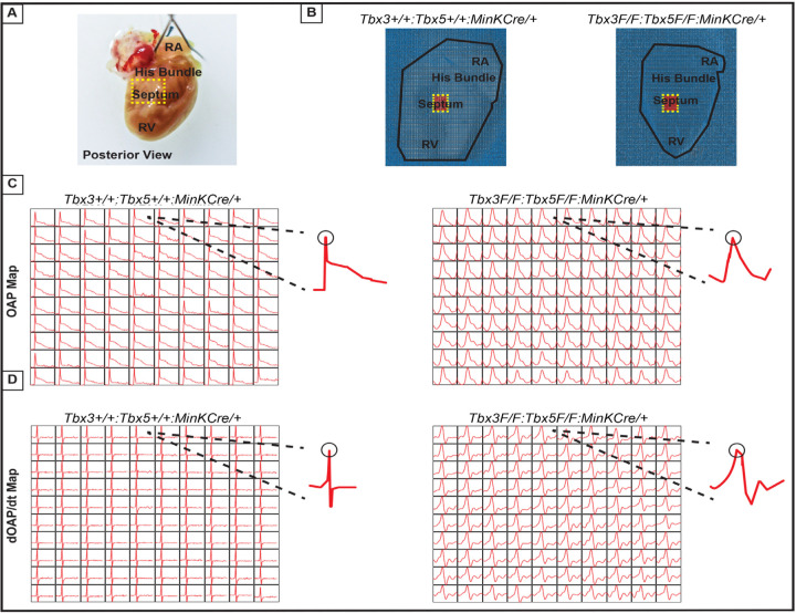
Figure 6. Ventricular optical action potentials (OAPs) distal from His bundle have only 1 OAP upstroke.
(A) Schematic of the posterior view of mouse heart with right ventricle (RV) free wall removed. (B) Representative 100x100 pixel OAP map recorded during sinus rhythm from Tbx3:Tbx5 double-conditional knockout mice and control littermates with RV free wall removed. The region of the working ventricular myocardium distal from the His bundle is highlighted in red. (C) Representative 10x10 pixel OAP map from the region distal to the His bundle. (D) Representative 10x10 dOAP/dt map from the region distal to the His bundle.

