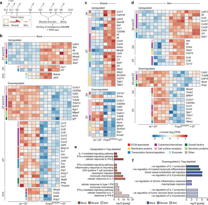
Fig. 5. Mo/MΦ from Treg-depleted mice show an increased pro-inflammatory transcriptional signature.
a Wildtype (wt) C57BL6/J and Foxp3DTR/GFP mice were treated with diphtheria toxin (DT) to deplete Tregs in Foxp3DTR/GFP mice, and injuries were performed in bone (critical-size cranial defects), muscle (quadriceps volumetric muscle loss defect) or skin (full-thickness dorsal skin wounds). b–f Mo/MΦ were sorted from the injured tissues at two different time points per tissue for RNA sequencing. Heat maps depicting standardised gene expression values of selected significantly upregulated and downregulated DEGs (FDR adjusted p value < 0.05) in Mo/MΦ from Treg-depleted bone (b), muscle (c) and skin (d) injuries, compared to the wildtype controls, including DEGs that were both shared and unique, across both time points per tissue (n = 3–5 mice/tissue per time point; individual replicates are shown). Colour key on the left of the heat map denotes the functional category of the genes. Gene ontology terms depicting enriched biological processes in the significantly upregulated (e) and downregulated (f) genes in the Treg-depleted tissues, from both time points combined (FDR < 0.01, adjusted by Benjamini–Hochberg correction). (Mo/MΦ: monocytes/macrophages). a Created with BioRender.com released under a Creative Commons Attribution-NonCommercial-NoDerivs 4.0 International license (https://creativecommons.org/licenses/by-nc-nd/4.0/deed.en).

