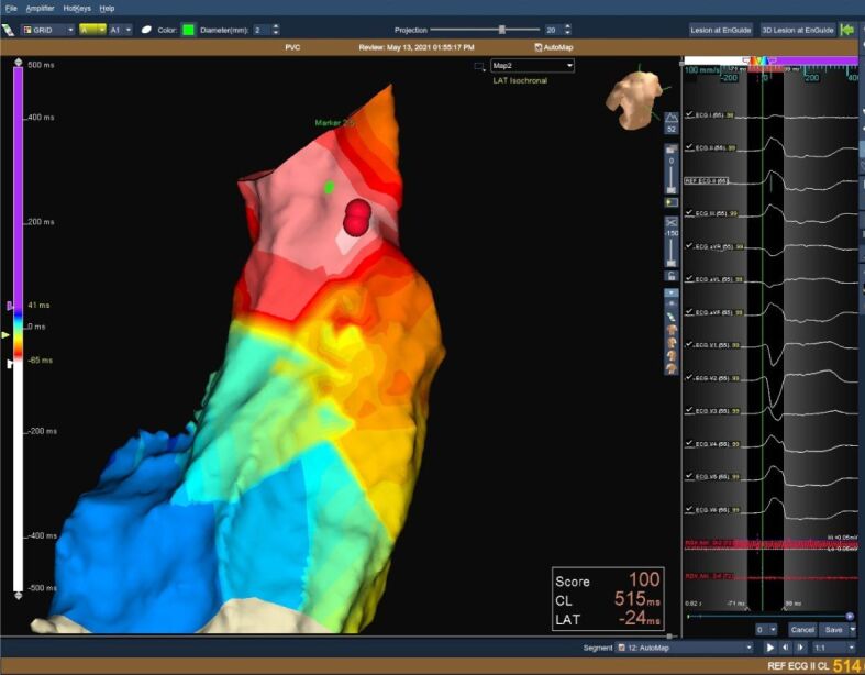
Figure 2.

3D reconstructed image of the RVOT (left panel) and 12-lead ECG (right panel) showing LBBB morphology PVCs with late transition in V4 R/S >1 in leads II, III, AVF. The presence of QRS notching and the lower R amplitude in the inferior leads is highly suggestive of a free wall origin. 3D: Three-dimensional; AVF: Augmented voltage foot; ECG: Electrocardiography; LBBB: Left bundle branch block; PVC: Premature ventricular contraction; RVOT: Right ventricular outflow tract
