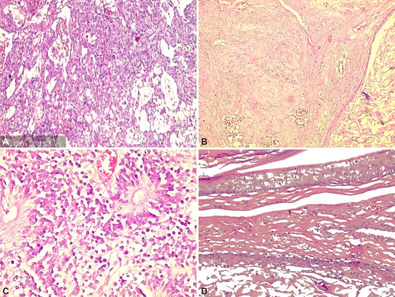
Figure 7.
Histopathology slides and description of Case No. 6: (A) Yolk sac tumor – anastomosing channels and variably sized cysts lined by primitive tumor cells with various amounts of clear to eosinophilic cytoplasm; (B) Germ cell tumor metastasis – after therapy was metastasized the MT, squamous epithelium, muscle tissue; (C) High-power view shows the immature teratoma component, with the presence of immature neural tissue; (D) Metastasis – MT with the presence of squamous epithelium. HE staining: (A and D) ×200; (B) ×100; (C) ×400. MT: Mature teratoma

