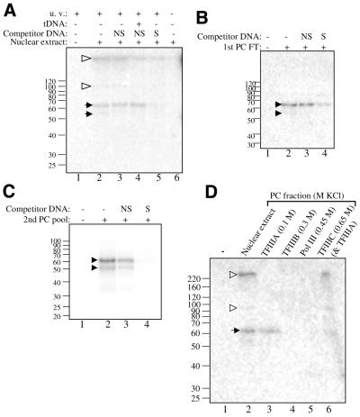
Figure 5.
The 59 KDa protein is detected in all TFIIIA-containing fractions. (A) Nuclear extract (73 µg of protein) with 12 µg of calf thymus DNA was cross-linked with 4600 c.p.m. of the +66 photoaffinity probe in a 30 µl reaction. The indicated reactions were treated with 150 ng of specific or non-specific competitor DNA. Lane 4 shows a reaction preincubated with 15 ng of a tRNAmet gene [EcoRI/HindIII digested pArabMet, 4 (22)]. Lane 6 shows a reaction that was not UV-irradiated. Black arrows identify AcTFIIIA, and open arrowheads show additional labeled polypeptides. (B) The first PC flow-through fraction (15 µg of protein) with 4 µg of calf thymus DNA was cross-linked with 4600 c.p.m. of the +66 photoaffinity probe in a 20 µl reaction. The indicated reactions were treated with 160 ng of specific or non-specific competitor DNA. (C) TFIIIA activity from the second PC column was cross-linked with the +86 photoaffinity probe essentially as described for the PC flow-through fraction. (D) PC column fractions (15 µg of protein per fraction) containing the separated Pol III factors with 4 µg of calf-thymus DNA were cross-linked with 2900 c.p.m. of the +66 photoaffinity probe. Above each lane, the KCl concentration applied to the column and the Pol III factor eluted are indicated.

