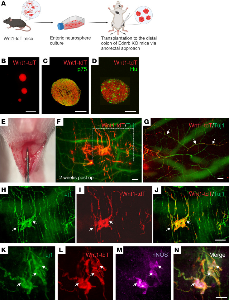
Figure 1. Transplant of Wnt1-tdT ENSCs to Ednrb-KO mice.
Schematic of experimental overview (A), including isolation of ENSCs from the gastrointestinal tract of Wnt1-tdT mice, their expansion as neurospheres (B), and subsequent transplantation into the aganglionic distal colons of Ednrb-KO mice via anorectal needle injection (A and E). Enteric neurospheres contain p75+ neural crest cells (C) and Hu+ neurons (D). Transplanted cells were observed 2 weeks following surgery (F), projecting fibers along host-derived Tuj1+ extrinsic nerves (G, arrows) and forming neo-ganglia (H–J, arrows) that contain donor-derived nNOS immunoreactive neurons (K–N, arrows). Scale bars: 50 μm (C, D, and K–N), 100 μm (F–J), and 200 μm (B).

