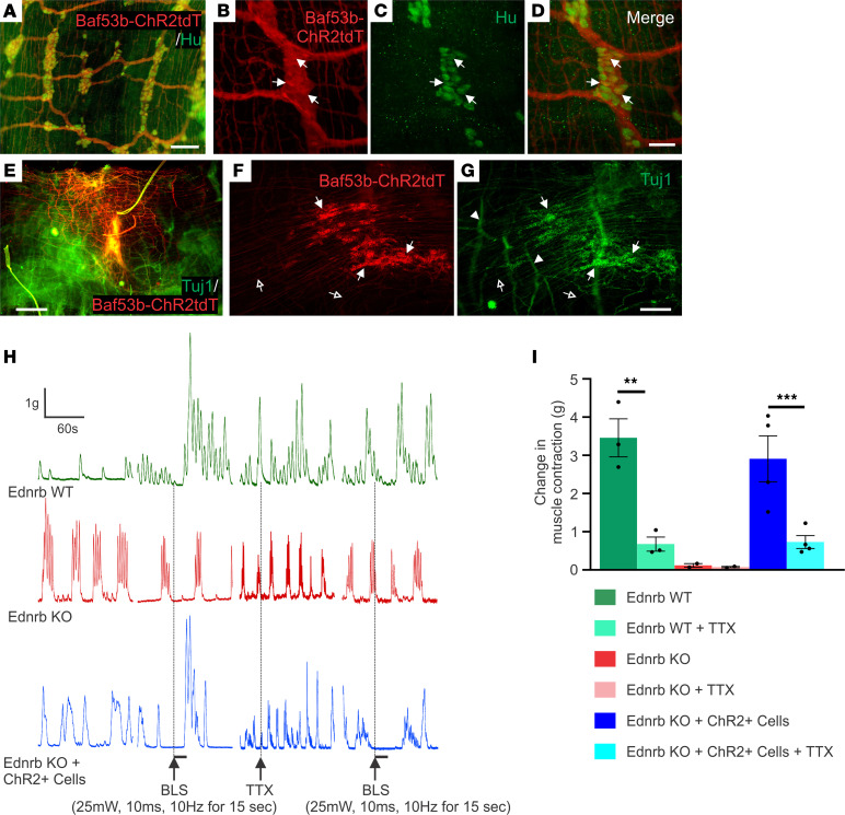
Figure 3. Optogenetics demonstrates neuromuscular connectivity between ENSCs and recipient aganglionic colon.
Immunohistochemical evaluation of ENS in the Baf53b-ChR2tdT mice confirmed that Hu+ enteric neurons express ChR2tdT (A–D, arrows). Two weeks after surgery, transplanted cells were visualized (E). High-power images show that transplanted cells form neuronal cell clusters (F and G, arrows) with projecting fibers (F and G, open arrows), and hypertrophic nerve bundles (F and G, arrowheads) within the aganglionic colon. Traces depict spontaneous contractions and smooth muscle responses to BLS (H). While Ednrb-KO and WT colon show no response to BLS, transplantation of ChR2-expressing ENSCs leads to robust smooth muscle contraction (I), which is significantly reduced by the addition of TTX (I). Scale bars: 50 μm (B–D), 100 μm (A), 200 μm (F and G), and 500 μm (E). All the values represent the mean of 2–4 animals for each group, repeated 2–3 times. Data are shown as the mean ± SEM. Statistical significance was determined by the 1-way ANOVA with a post hoc Tukey’s test. **P < 0.01 and ***P < 0.001 are statistically significant. BLS, blue light stimulation; ChR2, channelrhodopsin-2; TTX, tetrodotoxin.

