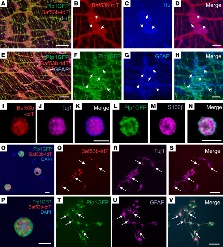
Figure 4. Isolation, expansion, and differentiation of ENSCs from Plp1GFP;Baf53b-tdT mice.
Plp1GFP;Baf53b-tdT mice, in which Baf53b/Hu+ neurons express tdT (A–D, arrows) and PLP1/GFAP+ glial cells express GFP (E–H, arrows) were used to isolate ENSCs and generate enteric neurospheres (I–P), which express markers for neurons (Tuj1;J) and glia (S100β;M). Upon dissociation and culturing on fibronectin, neurospheres give rise to neurons (Q–S, Tuj1, arrows) and glial cells (T–V, GFAP, arrows). Scale bars: 50 μm (B–D, I–K, and L–N), and 100 μm (A, E–H, and O–V).

