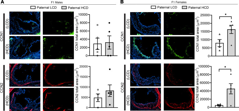
Figure 7. CCN1 and CCN2 proteins are elevated in the atherosclerotic lesions of F1 female LDL receptor–deficient descendants from high-cholesterol diet–fed sires.
Three-week-old male LDLR–/– mice were fed an LCD or HCD diet for 8 weeks before mating with control female LDLR–/– mice. Three-week-old F1 descendants were fed an LCD for 16 weeks. (A and B) Representative immunofluorescence images of CCN1 (green) and CCN2 (red) at the aortic root of F1 male (A) and female (B) offspring. The nuclei were stained with DAPI (blue). Scale bar: 100 μm. Quantification analysis of stating areas is displayed as indicated (n = 4–5, *P < 0.05, 2-sample, 2-tailed Student’s t test). All data are plotted as mean ± SEM.

