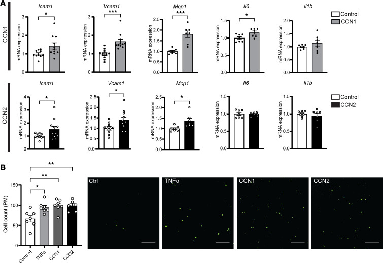
Figure 8. CCN1 and CCN2 proteins promote proatherogenic gene expression in endothelial cells in vitro.
(A) Human endothelial cells, HMEC-1 cells, were treated with 1 μg/mL CCN1 or CCN2 for 4 hours followed by total RNA isolation. The expression levels of indicated genes were analyzed by qPCR (n = 7–11, *P < 0.05, ***P < 0.001, 2-sample, 2-tailed Student’s t test). (B) HMEC-1 endothelial cells were pretreated with 50 ng/mL CCN1 or CCN2 or 10 ng/mL TNF-α for 24 hours before incubating with calcein acetoxymethyl–stained peritoneal macrophages isolated from LDLR–/– mice for 4 hours. Adhered cells were counted under a fluorescence microscope. Quantitative analysis of the adhered cells is displayed to the left of representative images (n = 6–7, *P < 0.05, **P < 0.01, 1-way ANOVA followed by Bonferroni’s multiple-comparison test). All data are plotted as mean ± SEM.

