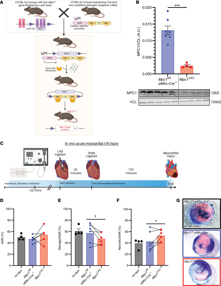
Figure 1. Loss of the MPC in murine hearts results in less myocardial salvage with more necrosis following I/R.
(A) The Mpc1 gene locus is targeted in C57Bl/6J mice by placing loxP sites in the introns flanking exons 3–5 of the Mpc1 genomic locus. Cardiomyocyte specificity was engineered by crossing Mpc1fl/fl mice with an Mhc driven tamoxifen inducible Cre. To induce deletion of Mpc1 (Mpc1CKO), mice were intraperitoneally injected with tamoxifen for 3 consecutive days (40 mg/kg) at 8 weeks old. (B) Western blot showing significant knockdown of MPC1 from whole hearts (2-tailed, Student’s t test) in Mpc1fl/fl-αmhc-cre–/–(n = 7) and Mpc1CKO (n = 4). Samples were run in parallel with one another, at the same time on separate gels blotting for MPC1 and VCL. (C) Schematic of in vivo myocardial I/R injury model. (D) Following I/R injury, the area at risk (AAR%) was nonsignificant (P = 0.13), indicating similar initial ischemic injury between tamoxifen treated Mpc1fl/fl-αmhc-cre–/– (46.67±2.45%, n = 6) and Mpc1CKO (54.33±5.42%, n = 6). (E and F) Within the area at risk, myocardial salvage (TTC: pink tissue staining) is significantly reduced (P = 0.04) in the Mpc1CKO (47.21±4.22%, n = 6) when compared with their paired tamoxifen treated Mpc1fl/fl-αmhc-cre–/– littermates (57.28±7.09%, n = 6). (G) Representative images of myocardial salvage and necrosis following in vivo I/R injury in nontamoxifen treated Mpc1fl/fl-αmhc-cre–/– (no-tam), and tamoxifen treated Mpc1fl/fl-αmhc-cre–/–, and Mpc1CKO mice. Paired t test was used for statistical analysis between the tamoxifen treated Mpc1fl/fl-αmhc-cre–/– and Mpc1CKO mice (D–F). There were no differences seen in the AAR, or myocardial salvage and necrosis between tamoxifen and nontamoxifen-treated (n = 4) Mpc1fl/fl-αmhc-cre–/– animals (2-tailed, Student’s t test). *P < 0.05, ***P < 0.001. Values are represented as mean±SEM. BioRender was used to make panels A and C.

