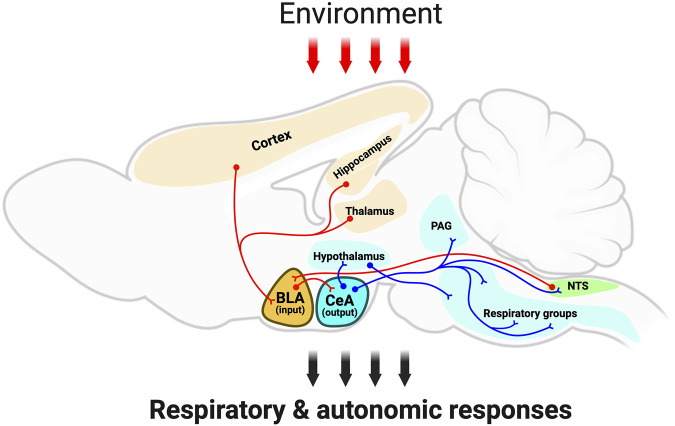
FIGURE 1.
Schematic outlining the amygdala integratory function and neuronal anatomical connectivity. The schematic, red-colored neurons depict the sensory information from several brain regions, such as brainstem (i.e., NTS), cortex, thalamus, and hippocampus to the amygdala; and the red arrows depict sensory information from the environment targeting the amygdalar-neuroanatomical network. Note that the basolateral amygdala subdivision is a gating center that receives sensory information, which is further transmitted to the central amygdala. Then, the central amygdala sends projection neurons (blue-colored neurons) to hypothalamic centers, periaqueductal grey, as well as brainstem centers. This amygdalar-neuroanatomical framework might modulate the ongoing respiratory motor activity for respiration and airway-protective behaviors (e.g., cough, sneeze, laryngeal adduction, and swallow). Abbreviations: BLA, basolateral amygdala; CeA, central amygdala; Hypo, hypothalamus; PAG, periaqueductal gray; NTS, nucleus of the tractus solitarius; RG, respiratory groups (in the brainstem). Created with BioRender.com.

