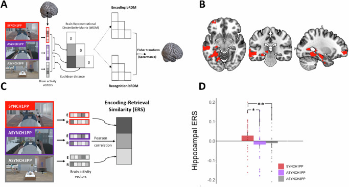
Fig. 3. Left hippocampal ERS is higher under visuomotor and perspectival congruency.
A RSA identified brain regions with the same differential pattern between conditions during encoding and recognition sessions in Experiment 2. B RSA identified four regions: left hippocampus, visual cortex, left middle temporal, and left frontal superior orbital gyrus (permutation test, Npermutations = 1000, p < 0.05, cluster size> 500). C ERS was computed for the four regions identified by RSA, by applying a Pearson correlation between the voxel activity at encoding and at recognition in Experiment 2. D Hippocampal ERS is significantly higher under visuomotor and perspectival congruency (SYNCH1PP, red) compared to the two other conditions. Error bars represent the standard error of the mean. * and ** indicates significance level with p-value < 0.05 and <0.01 respectively. ERS Encoding recognition similarity score. RSA Representational Similarity Analysis, N = 24.

