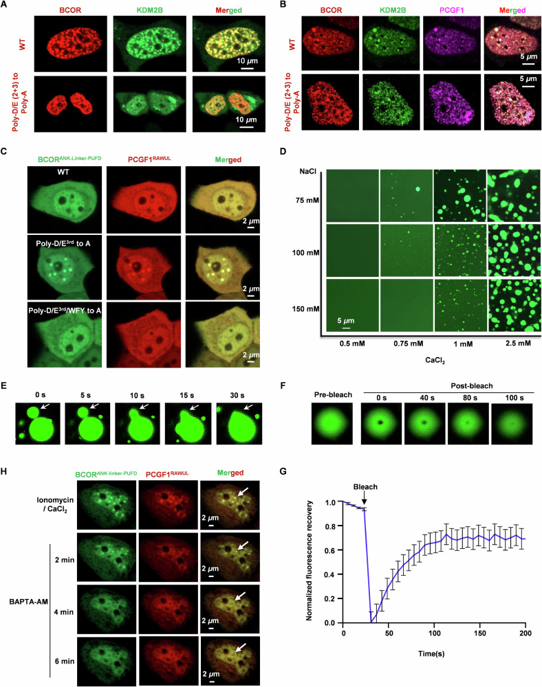
Fig. 5. Liquid-liquid phase separation of BCORANK-linker-PUFD/PCGF1RAWUL hetero-dimer.
A Co-localization analysis of BCOR-mCherry with KDM2B-EGFP in live HeLa cells. B Co-localization analysis of BCOR-mCherry, PCGF1-ECFP and KDM2B-EGFP in live HeLa cells. C Observation of condensates for BCORANK-linker-PUFD (wild type or mutant)/PCGF1RAWUL in HeLa cells. WFY indicates the 5 aromatic residues on linker of BCOR, including W1598, F1600, Y1601, Y1614 and Y1633. D Effect of CaCl2 and NaCl on the phase separation of BCORANK-linker-PUFD/PCGF1RAWUL hetero-dimer with concentration of 60 μM. E Representative fusion event of BCORANK-linker-PUFD/PCGF1RAWUL droplets. F In vitro FRAP analysis of the droplet of BCORANK-linker-PUFD/PCGF1RAWUL. G Normalized FRAP recovery curves for BCORANK-linker-PUFD-EGFP/PCGF1RAWUL droplet in (F). Data are mean ± SEM at each time point and combined data from three independent repeats. H Plasmids expressing BCORANK-Linker-PUFD or PCGF1RAWUL were co-transfected into HeLa cell. Before imaging for LLPS, cells were pretreated with 10 μM ionomycin and 5 mM CaCl2 for 2 h. To observe the effect of calcium-chelator BAPTA-AM on the ionomycin induced LLPS, culture containing 10 μM ionomycin and 5 mM CaCl2 was discarded, the cell was washed with PBS, then culture containing 10 μM BAPTA-AM was added to the cell.

