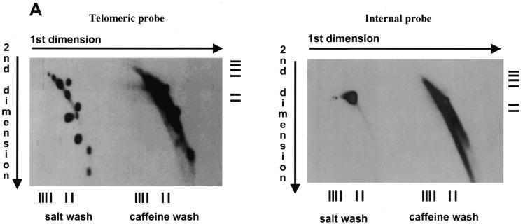
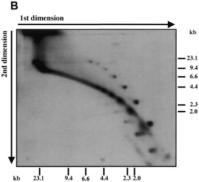
Figure 3.
Telomeric fragments elute from BND–cellulose as duplex DNA and are present in the undigested preparations of C.parapsilosis DNA. (A) Total DNA of C.parapsilosis digested with BglII was loaded on a BND–cellulose column and eluted first with 0.8 M NaCl (salt wash) followed by 1.8% caffeine in 1 M NaCl (caffeine wash). The eluted DNA was separated by neutral-neutral 2-D electrophoresis as in Figure 1, and hybridized with a radiolabeled 738 bp telomeric tandem repeat unit (telomeric probe) or a 2.9 kb BglII fragment of mtDNA of C.parapsilosis internal to the telomere (internal probe). The bars represent DNA marker fragments of 23.1, 9.4, 6.6, 4.4, 2.3 and 2.0 kb, respectively. (B) Undigested total DNA of C.parapsilosis (not subjected to BND–cellulose chromatography) was separated by neutral-neutral 2-D electrophoresis and hybridized with a radioactively-labeled 738 bp telomeric tandem repeat unit probe.


