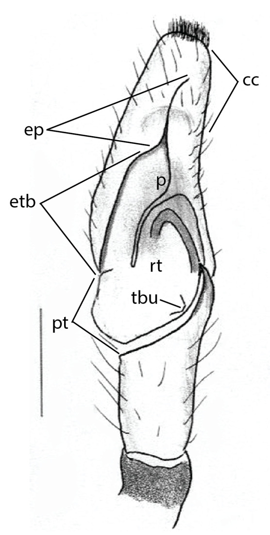
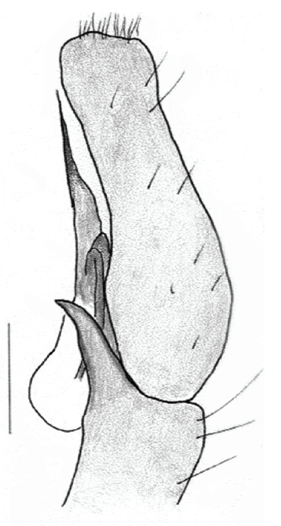
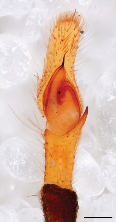
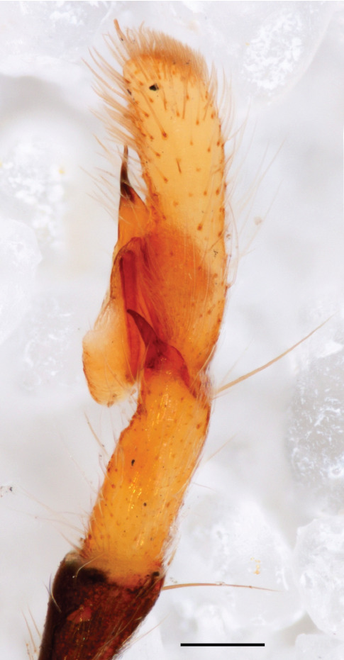
Annotated illustrations and photographs of male pedipalp in Chrysillalauta Thorell, 1887
Figure 1a.

Chrysillalauta Thorell, 1887, left male pedipalp, ventral view cc cymbium cap ep embolus proper etb embolar tegular branch p distal projection of embolar tegular branch beyond retrolateral lobe of tegulum excluding embolus proper pt proximal lobe of tegulum rt retrolateral lobe of tegulum tbu tegular bump. Scale bar: 0.3 mm
Figure 1b.

Chrysillalauta Thorell, 1887, left male pedipalp, retrolateral view. Scale bar: 0.3 mm
Figure 1c.

Chrysillalauta Thorell, 1887, ventral view, CM 15726, scale bar 0.2 mm
Figure 1d.

Chrysillalauta Thorell, 1887, retrolateral view, CM 15726, scale bar 0.2 mm
