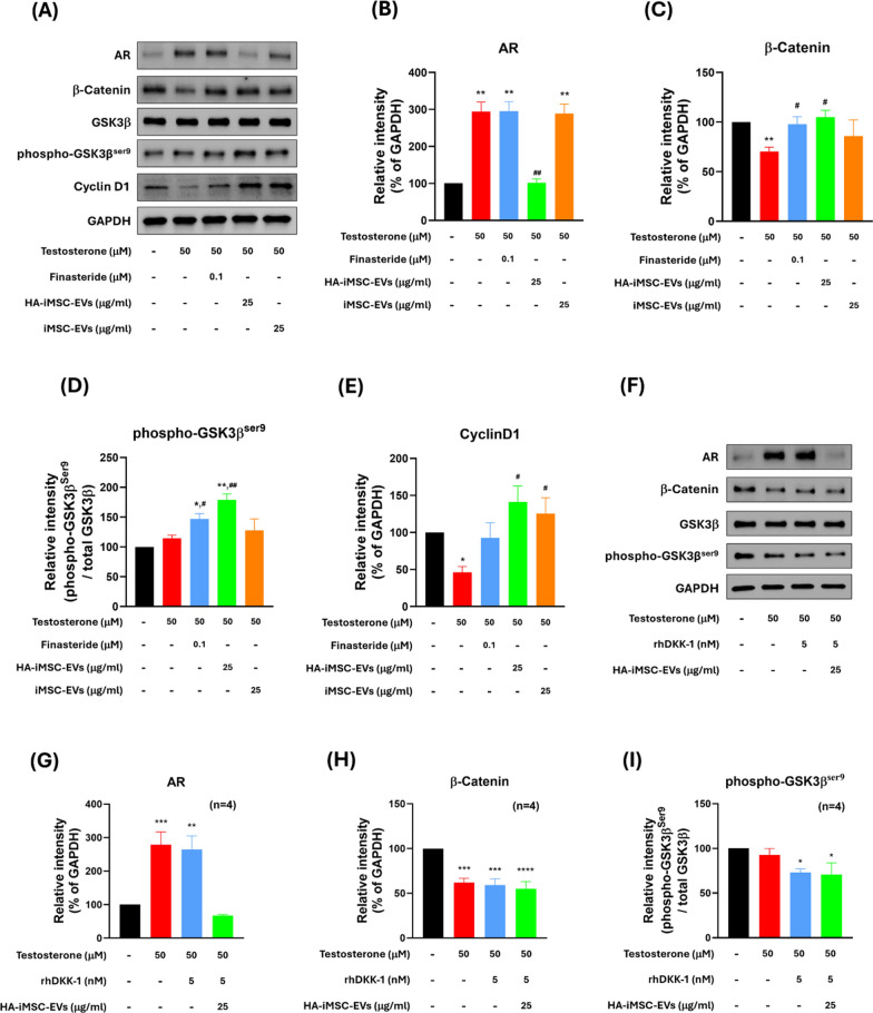
Fig. 3.
HA–iMSC–EVs regulated AR-related Wnt/β-catenin signaling and proliferation in HFDPC. A–E The protein expression was detected with 50 μM testosterone in the presence of 100 nM finasteride, 25 μg/mL HA–iMSC–EVs or 25 μg/mL iMSC–EVs for 24 h in HFDPC. A A typical western blotting result showing that HA–iMSC–EVs reduced AR expression and alleviated testosterone-induced alopecia. B–E The expression of each protein was normalized against that of GAPDH (N = 4). Uncropped western blot images are shown in Additional file 1: Fig. S3. F–I Inhibition of anti-AGA effects of HA–iMSC–EVs by rhDKK-1. HDFPCs were treated with 50 μM testosterone in the presence of 5 nM rhDKK-1 or 25 μg/mL HA–iMSC–EVs for 24 h. The expression of AR, β-catenin, and phospho-GSK3βser9 was normalized against that of GAPDH (N = 4). Uncropped western blot images are shown in Additional file 1: Fig. S4. The protein bands were measured by the ImageJ software and plotted as mean ± SEM. *p < 0.05, **p < 0.01 versus control group. #p < 0.05, ##p < 0.01 versus testosterone-only group. EV, extracellular vesicle; HA, hyaluronic acid; HFDPC, hair follicle dermal papilla cell; iMSC, induced pluripotent stem cell-derived mesenchymal stem cell

