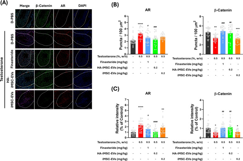
Fig. 6.
Immunofluorescence detection of androgen receptor (AR) and β-catenin in the hair follicle of AGA mice. A Representative images of AR and β-catenin expression in AGA skin tissues. The outline of the hair follicle is indicated by a dotted line. Red and green colors indicate AR and β-catenin, respectively. Scale bars = 25 μm. B The AR-positive and β-catenin-positive cells were counted in the form of puncta in the hair follicle area. C Quantification of the relative expression of AR and β-catenin in the hair follicle area. B, C The symbols represent the value of all individual hair follicles counted in five mice. Quantified values from each hair follicle are indicated by gray symbols in B and C. *p < 0.05, **p < 0.01, ***p < 0.001, ****p < 0.0001 versus vehicle control mice. #p < 0.05, ##p < 0.01, ###p < 0.001, ####p < 0.0001 versus testosterone-only group. AGA, androgenetic alopecia

