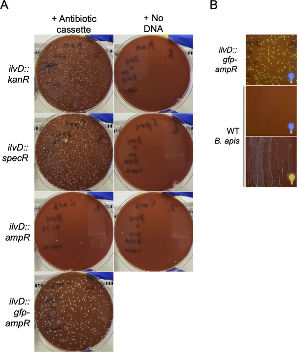
Fig 6.
The B. apis genome can be engineered with a lightweight approach. (A) Plates demonstrating that ilvD can be knocked out in B. apis. Col-B + kan agar plates inhibit WT B. apis growth (top right) and select for insertion of ilvD::kanR (top left). Col-B + spec agar plates inhibit WT B. apis growth (middle-top right) and select for insertion of ilvD::specR (middle-top left). Col-B + carb agar plates inhibit most WT B. apis growth (middle-bottom right; a few spontaneously resistant mutants colonies are present) and select for insertion of ilvD::ampR (middle-bottom left) and ilvD::gfp-ampR (bottom left). (B) Plates illustrating successful ilvD::gfp-ampR transformation into B. apis. ilvD::gfp-ampR colonies fluoresce green in front of a blue light transilluminator (top), whereas WT B. apis (bottom) does not not fluoresce (middle). See panel A (bottom left) for ilvD::gfp-ampR colonies imaged under ambient light. Blue light bulb: plate imaged in front of blue light; yellow light bulb: plate imaged under ambient light.

