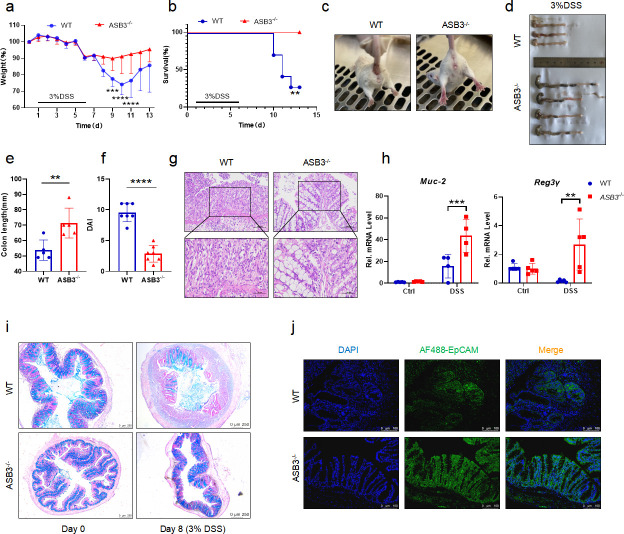
Fig 2.
ASB3 deficiency provides protection against DSS-induced colitis. (a and b) Body weight (a) and survival (b) of conventionally raised wild-type and ASB3−/− mice treated with 3% DSS (above horizontal axes) (WT, n = 7; ASB3−/−, n = 7); the results are presented relative to initial values, set as 100% (throughout). (c) Representative images of diarrhea/bloody stools observed on day 6. (d and e) Colon length on day 8. (f) DAI scores were determined on day 6. (g) Representative images of pathological H&E-stained colon sections collected on day 8. Scale bar, 100 or 50 µm. (h) The mRNA expression levels of Muc-2 and Reg3γ were determined by qPCR assay. (i) Representative images of Alcian Blue staining of colon tissues of WT and ASB3−/− mice. Scale bar, 250 µm. (j) IF for EpCAM in the colon; representative image of three mice/genotype. Scale bar, 100 µm. Differences between groups in panels e and f were determined by unpaired Student’s t test, in panels a and h by one-way ANOVA, and in panel b by log-rank test.

