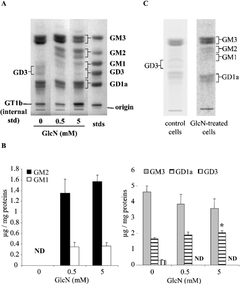
Figure 4. Effect of GlcN on ganglioside profile.
(A, B) RMC were treated with GlcN (0.5 and 5 mM) for 48 h and then collected. Gangliosides were extracted, purified and then analysed by HPTLC and resorcinol staining. Representative HPTLC (A) and densitometric quantification (B) are shown. Results are expressed as μg of gangliosides/mg of protein and are the means±S.E.M. for four independent experiments each performed in duplicate. Under control conditions, ganglioside quantities were 4.62±0.37, 1.65±0.11 and 0.30±0.05 μg/mg of protein for GM3, GD1a and GD3 respectively. ND, not detectable. *P<0.05 versus control. (C) RMC were labelled for 48 h in the presence of 0.2 μCi/ml [14C]GlcN for control cells and 2 mM GlcN at a specific activity of 0.5 mCi/ml for treated cells. At the end of incubation, cells were washed extensively and gangliosides were extracted, purified and separated by HPTLC as described in the Experimental section. Radioactivity associated with gangliosides was then analysed by autoradiography of the HPTLC plates.

