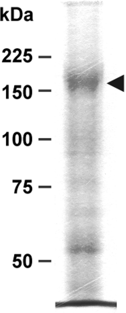
Figure 7. P-gp as revealed by electrophoresis of DRM-containing fraction.

Proteins (10 μg) from fraction F3 were deposited on an SDS/8% polyacrylamide gel and run as described in the Materials and methods section. The gel was stained with Coomassie Blue. The arrowhead points to the band corresponding to P-gp.
