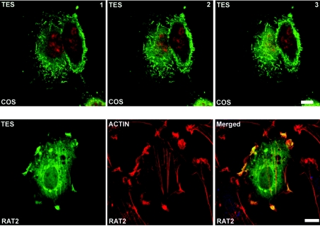
Figure 6. Immunofluorescence studies of Tes overexpressed in COS-7 and Rat2 cells.
COS-7 cells (upper panel) and Rat2 cells (lower panel) were transiently transfected with the plasmid pEF-DEST51-Tes. The upper panel corresponds to three acquisitions of the same COS-7 cells from the basal surface (image 1) to the middle part and the top of the cells (images 2 and 3, respectively; Z scaling: 0.39 μm). Tes (pseudo-coloured in green) was stained with anti-V5 mAb and Alexa Fluor™ 546 goat anti-mouse. Nuclei (pseudo-coloured in red) were counterstained with TO-PRO 3. In the lower panel (Rat2 cells), Tes was stained with anti-V5 antibody and Alexa Fluor™ 488 goat anti-mouse. Tes (pseudo-coloured in green) co-localizes with F-actin, labelled with Alexa Fluor™ 568 phalloidin and pseudo-coloured in red (see yellow staining in merged image). Scale bar=10 μm.

