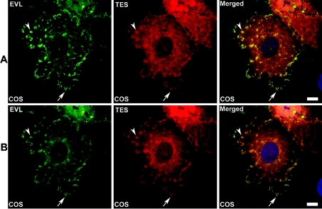
Figure 8. Immunofluorescence studies of EVL and Tes in COS-7 cells.
COS-7 cells transiently co-transfected with the plasmid DEST-EVL and the plasmid pEF-DEST51-Tes were stained using respectively anti-EVL polyclonal antibodies and Alexa Fluor™ 488 goat anti-rabbit (EVL pseudo-coloured in green) and anti-V5 mAb and Alexa Fluor™ 546 goat anti-mouse (Tes pseudo-coloured in red). Nuclei (pseudo-coloured in blue) were counterstained with TO-PRO 3. Filopodia and focal adhesions are pointed out by arrow and arrowhead, respectively. In (A), image acquisition was performed at the plane of coverslip contact and (B) corresponds to the upper optical section of the same cells (Z scaling: 0.32 μm). Scale bar=10 μm.

