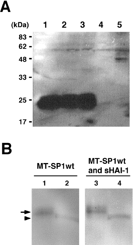
Figure 5. Western-blot analysis of the NTF of a recombinant MT-SP1/matriptase expressed in COS-1 cells.
(A) Representative Western blot showing the 22 kDa band derived from the recombinant MT-SP1/matriptases expressed in COS-1 cells. Transfection was performed when cells cultured on 100 mm dishes reached approx. 90% confluence. Samples were separated by SDS/PAGE (12% polyacrylamide) under reducing conditions, and Western-blot analysis was performed using Tmc172. Lane 1, sample from COS-1 cells transfected with pcMT-SP1MycHis (25 μg); lane 2, sample from COS-1 cells transfected with pcMT-SP1wt (12.5 μg) and pSecTagHygro vector (12.5 μg); lane 3, sample from COS-1 cells transfected with pcMT-SP1wt (12.5 μg) and pSecHAI-1 (12.5 μg); lane 4, sample from COS-1 cells transfected with pSecHAI-1 (12.5 μg) and pcDNA3.1(+) vector (12.5 μg); lane 5, sample from COS-1 cells transfected with pcDNA3.1(+) vector (25 μg). (B) Representative Western blot showing the cell-surface localization of the NTF. Transfection was performed when cells cultured on 100 mm dishes reached approx. 90% confluence. Samples were separated by SDS/PAGE (16% polyacrylamide) under reducing conditions, and Western blots were probed with Tmc172. Lanes 1 and 2, the eluate and flow-through fractions respectively from streptavidin–agarose-precipitated proteins of COS-1 cells transfected with pcMT-SP1wt (12.5 μg)+pSecTagHygro vector (12.5 μg); lanes 3 and 4, eluate and flow-through fractions respectively from streptavidin–agarose-precipitated proteins of COS-1 cells transfected with pcMT-SP1wt (12.5 μg)+pSecHAI-1 (12.5 μg). Note that the biotinylated NTF (indicated by an arrow) migrated more slowly than the NTF in the flow-through fraction (indicated by an arrowhead) because of the addition of the sulpho-NHS-SS-biotin spacer.

