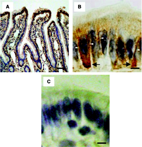
Figure 8. Immunostaining of rat jejunum with Tmc172.
(A) Staining of the rat jejunum along the crypt-villus axis. (B) Staining on the villus tip. Staining on the lateral membranes and in the cytosol close to the basal membrane is indicated by open arrowheads and arrows respectively. (C) Staining on the villus tip when Tmc172 was preadsorbed with the antigenic peptide at a concentration of 1 mM. Counterstaining was with haematoxylin. Scale bars: 200 μm for (A) and 10 μm for (B, C).

