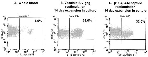
FIG. 3.
Flow cytometric analysis of MmL-3 T cells stained with Mamu A*01/p11C tetramer complex. A fresh blood sample (A), 2-week CTL culture restimulated with Vac-SIVgag (B), and 2-week CTL culture restimulated with p11C peptide (1 μg/ml) (C) from MmL3 were stained with the tetramer complex and anti-CD3 and anti-CD8 monoclonal antibodies. The events were collected and sequentially gated for lymphocyte population (forward versus side scatter) and CD3+ CD8+ population. The percentages shown in the figure indicate the tetramer staining-positive population within the CD3+ CD8+ lymphocytes.

