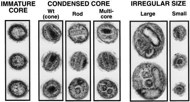
FIG. 2.
Electron microscopic classification of virion morphology. Three typical examples of each type of morphology (except irregular large) are shown. The upper two virions in each category are typical longitudinal cross-sections, and the lower virion in each category is a typical latitudinal cross-section. Virus was pelleted from clarified supernatants of 293T cells 72 h after transfection with mutant or WT plasmid DNA. Images are digital and were scanned from electron micrographs at a magnification of ×90,000.

