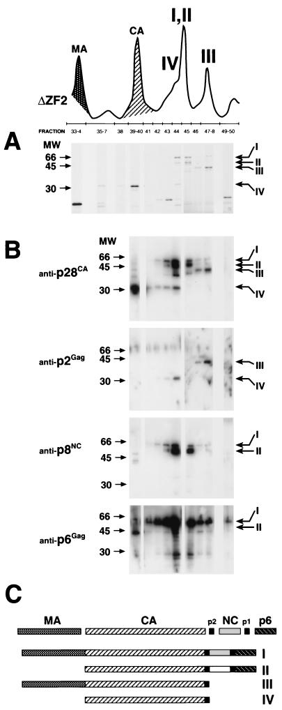
FIG. 7.
SDS-PAGE and immunoblot analysis of incomplete Gag polyprotein processing. (A) A section of the chromatograph from Fig. 6 (ΔZF2) showing an example of the NC mutant, ΔZF2, RP-HPLC, and numbered protein fractions that were fractionated by SDS-PAGE and visualized with Coomassie stain. Four distinct proteins (I to IV) were noted in peaks more prominent in the NC mutant than in the WT SIV(Mne) chromatographs. (B) Immunoblot analyses of fractions for anti-p28CA, anti-p2Gag, anti-p8NC, and anti-p6Gag antibodies. Molecular markers are shown on the left; the positions of labeled proteins I to IV are indicated on the right. Note that the p2Gag antibody only recognizes a cleaved or C-terminal epitope. (C) Identity of incompletely processed Gag proteins I to IV based on Coomassie, immunoblot, and partial amino acid sequence analyses. The same Gag polyproteins I to IV were observed in all NC mutants, only in different amounts.

