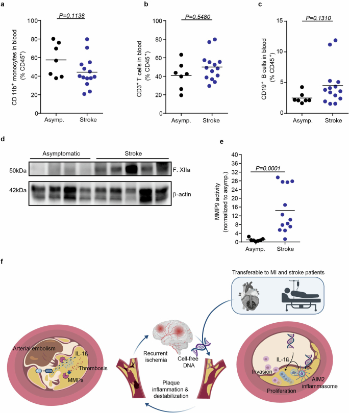
Extended Data Fig. 10. Blood leukocyte counts do not differ between stroke and asymptomatic patients with high-grade atherosclerosis.
a–c. Flow cytometry analysis of blood from asymptomatic patients or stroke patients showing the percentage of monocytes (CD11b+), T cells (CD3+) and B cells (CD19+) out of total leukocytes (CD45+) (U test, asymptomatic patients, n = 7; symptomatic patients, n = 13). d. Representative immunoblot from asymptomatic and stroke patients for F. XIIa and β-actin (Quantification can be found in Fig. 4i). e. Quantification of MMP9 activity normalized to the activity in asymptomatic patients (Representative image shown in Fig. 4h). Raw membrane images of immunoblots with cropping indication can be found in Supplementary Fig. 1q. f. Overview schematic: Stroke leads to the release of NETosis-derived cell-free DNA activating the AIM2 inflammasome and subsequent secretion of IL-1β. The release of IL-1β drives MMP expression in atherosclerotic plaque, leading to fibrous cap destabilization. The fibrous cap rupture initiates the activation of the intrinsic coagulation cascade resulting in atherothrombosis and subsequent arterio-arterial embolism with secondary brain infarctions.

