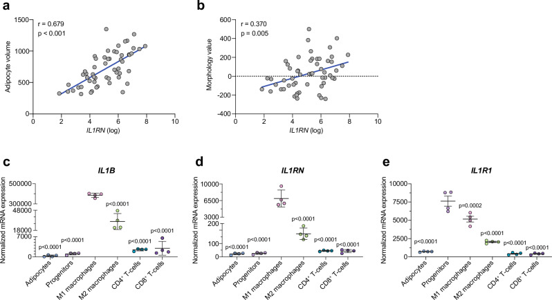
Fig. 3. Reduced IL-1 signaling correlates to hypertrophic adipocytes, and progenitors express the highest levels of IL1R1 in human scWAT.
a, b Pearson correlation of human scWAT IL1RN expression and adipocyte volume (a) or morphology value (b) where positive and negative values indicate WAT hypertrophy and hyperplasia, respectively (n = 56). Two-tailed p-values. c-e Expression of IL1B (c), IL1RN (d) and IL1R1 (e) in adipocytes and FACs-sorted stromal vascular cell populations from human scWAT (n = 6 donors, pooled 3 and 3 and processed in duplicates). Statistical analyses by one-way ANOVA and Dunnett’s multiple comparisons test compared to M1 macrophages (c, d) or progenitors (e). Line and error bars represent mean ± SEM. Source data are provided as a Source Data File.

