
Fig. 2.
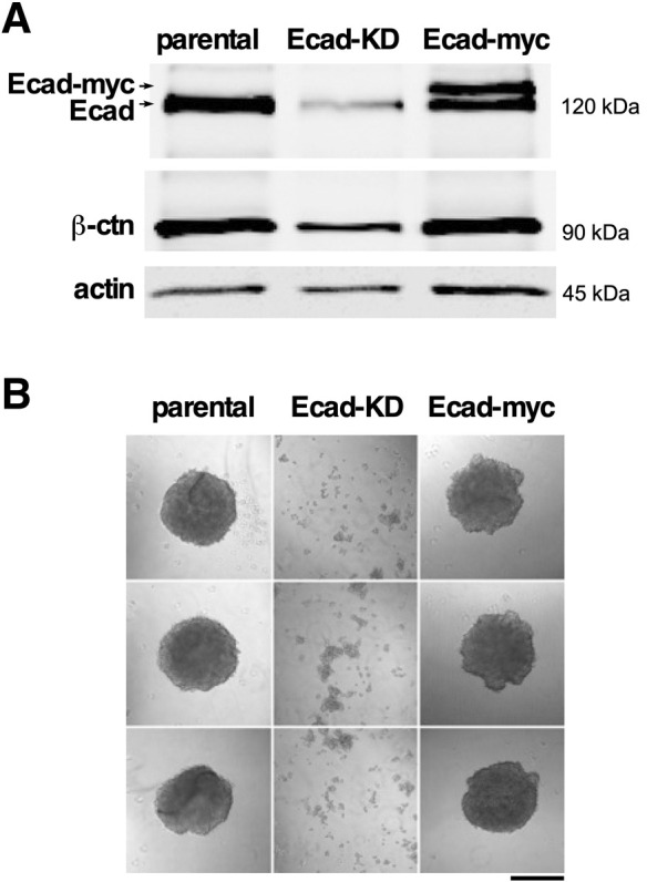
Depletion of E-cadherin in 4T1 cells leads to a reduction in β-catenin and loss of E-cadherin function. (A) Equivalent amounts of NP40 lysates of 4T1 cells (parental), Ecad-KD and Ecad-myc cells were loaded onto a SDS page gel. The proteins were size separated, transferred to membranes and incubated with antibodies to the proteins indicated. The primary antibodies were recognized by fluorescently labeled secondary antibodies and the membrane was imaged using a LI-COR blot system. The upper band in lane 3 corresponds to the anticipated size of myc-tagged E-cadherin. Blots are representative of two independent experiments. (B) Parental, Ecad-KD and Ecad-myc cell spheroids were recovered by pipetting (see Materials and Methods) and plated in 35 mm dishes for immediate photography. Knockdown of E-cadherin severely compromised the spheroid stability, indicating strong downregulation of cell-cell adhesion. Images are representative of two independent experiments. Scale bar: 500 μm.