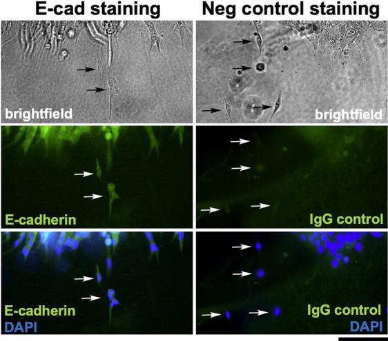
Fig. 7.

E-cadherin is retained in single cells and clusters of cells emerging from 4T1 cell spheroids embedded in 3D collagen. The spheroid depicted in Fig. S4 and Movies 2 and 3 was fixed after the 40-h time-lapse experiment and stained with an anti-E-cadherin antibody. Another spheroid from a parallel experiment was fixed and stained with a mouse IgG isotype negative control antibody. For both samples, cell nuclei were visualized with DAPI, and the edges of the spheroids were imaged by brightfield and fluorescence microscopy. Arrows point to single cells or small groups of cells that have emerged from the spheroids. These results demonstrate that cells and cell clusters invading into the 3D collagen matrix either retained E-cadherin expression throughout the invasion process or rapidly re-expressed E-cadherin upon dissociation from the spheroid. Images are from one experiment in which spheroid invasion was monitored by time-lapse imaging followed by immunostaining. Scale bar: 100 μm.
