
Fig. 8.
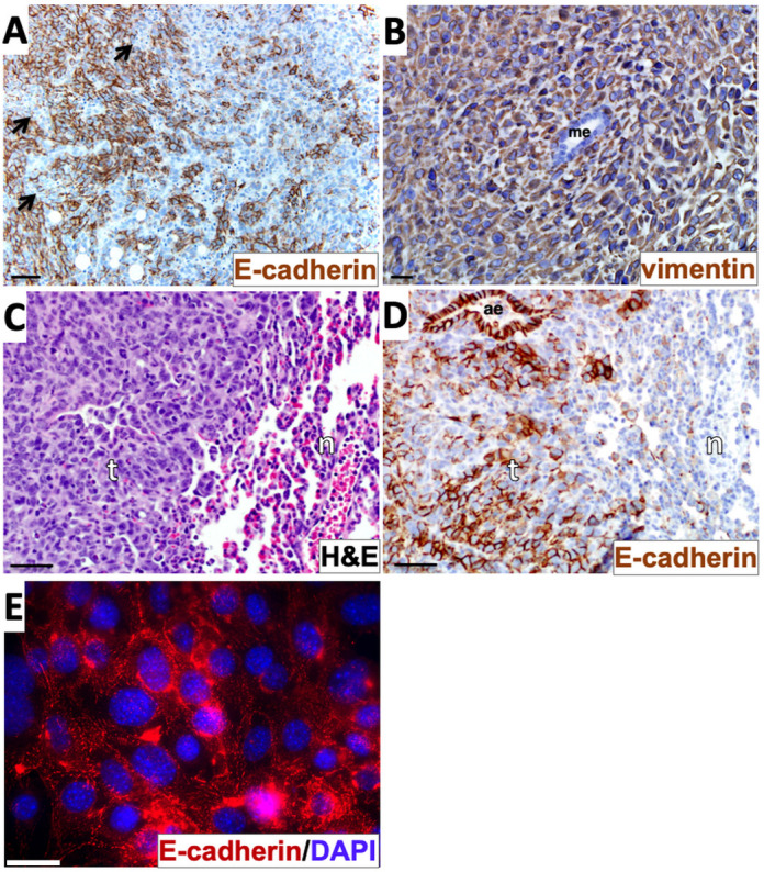
Evidence of a hybrid epithelial/mesenchymal phenotype and epithelial-mesenchymal transition-like events in the 4T1 model. (A) Primary tumor tissue was stained with E-cadherin immunohistochemistry (brown) and counterstained with Hematoxylin. Arrows indicate E-cadherin-negative zones within an overall E-cadherin-positive sector of the tumor. (B) Primary tumor tissue was stained with vimentin immunohistochemistry (brown) and counterstained with Hematoxylin. (C,D) Tissue from lung metastatic tumors were stained with Hematoxylin and Eosin (H&E) (C) or with E-cadherin immunohistochemistry and Hematoxylin counterstain (D). (E) Cells explanted from a 4T1 metastatic lung tumor as described in the Materials and Methods were immunostained with anti-E-cadherin (red) and labelled with DAPI (blue). Images in A-D are representative of three different tumor sections taken from one in vivo experiment. The image in E represents cells from one lung metastasis explanted in vitro. ae, airway epithelium; n, normal lung tissue; me, mammary epithelium; t, tumor tissue. Scale bars: 50 µm (A,C,D); 20 µm (B); 30 μm (E).